OUR SERVICES
We are the competence center for nuclear diagnostics and therapy
Therapies in Nuclear Medicine

Many functions of various organs can be examined by scintigraphy. The prerequisite for this is that, depending on the organ to be examined, the radioactive substance intended for this purpose is selected. The amount of radioactivity supplied to you usually causes a low radiation exposure. In children, a lower bodyweight-related activity is generally chosen.
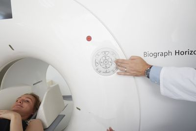
Learn morePET/CT
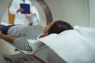
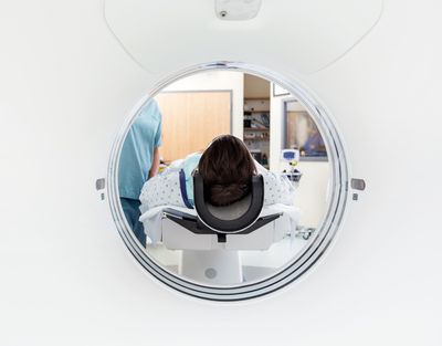
The examination is similar to a normal scintigraphy with a previous injection into a vein. Subsequently, images of different body sections with a special camera, the so-called PET scanner, a kind of open tube, made. Depending on the clinical question and the body sections to be examined as well as the radioactive medicine used, the duration of the examination is approx. 1-2 h.
Learn more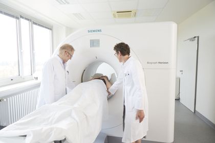
Schilddrüsenerkrankungen

Nuclear medicine mainly offers a thyroid consultation, which holds all technical examinations as well as blood analysis for thyroid diagnostics in addition to the consultation
Learn morerenal scintigraphy
Nuclear medical kidney diagnostics are used to visualize the blood flow, various kidney functions and urinary outflow. As a rule, a weakly radioactive substance is administered intravenously, which is then excreted via the kidneys. Measurements of blood radioactivity (blood samples) are then made and an image sequence (scintigraphy) made.
Learn more
Myokardszintigraphie
There is myocardial scintigraphy (myocardial perfusion scintigraphy) and radionuclide ventriculography (RNV), which is less frequently used today. How does the diagnosis of the heart work? Both examinations consist of two parts: a resting and a load investigation. In myocardial scintigraphy, the circulation of the heart muscle under stress is first ...
Learn morebrain scan
With the help of perfusion scintigraphy, it can be determined whether relevant circulatory disorders are present in the brain. This study may be required for symptoms such as prolonged dizziness, episodes of amnesia, cognitive or motor disorders.
Learn more
Somatostatin receptor scintigraphy in pancreatic cancer (pancreatic cancer)
If you have a neuroendocrine tumor found, then this scintigraphy is regularly used in post-operative care to ensure that any suspicious tissue has been removed. Also in the further aftercare, this investigation plays a major role. Rarely, it is used as a primary diagnosis before the start of therapy, here the PET / CT with somatostatin receptor ligands plays an important role.
Learn morelung scintigraphy
In pulmonary scintigraphy, various sub-functions such as blood circulation, ventilation and lung cleansing can be examined. Most often, evaluation of pulmonary blood flow to exclude obstruction of a pulmonary vein (pulmonary embolism) is necessary.
Learn more
Scintigraphy of the digestive system
In some diseases of the rheumatic type, eg collagenoses, there may be a limitation of the mobility of the esophagus, which can be felt, inter alia, by dysphagia. Another disease that accompanies a passageway disorder of the upper digestive tract is the "dumping syndrome".
Learn moreLymphknotenszintigraphie
Sentinel is called in German guards. Sentinel lymph node scintigraphy searches for the lymph node closest to the tumor. This is very important because of the large differences in the lymphatic drainage from person to person and also helps to avoid the unnecessary removal of several healthy lymph nodes.
Learn moreEntzündungsszintigraphie
In the inflammatory scintigraphy, a focus of inflammation is sought in the body. This may be the case in a fever or fever episode, but the cause has not been found. In addition, inflammatory changes after surgery (eg introduction of foreign bodies in artificial hip or knee joints) can be detected or excluded.
Learn moreBone scan
The bone metabolism can be disturbed both in benign (eg rheumatism) and in malignant diseases (eg bone cancer). To examine the bone metabolism, a weakly radioactive substance, which accumulates in particular on the bone surface, is injected into a vein of the arm.
Learn more
Nebennierenszintigraphie